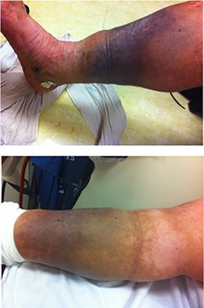
Deep Vein Thrombosis (DVT)
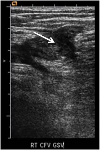
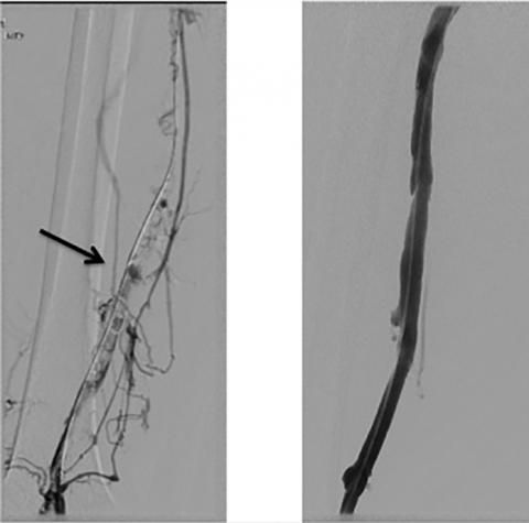
Catheter directed treatment of an acute DVT. The ultrasound shows a new clot in the deep vein of the leg (arrow). The first dye study shows extensive clot in the deep vein of the lower thigh, with dye trying to go through alternate pathways to bypass the blockage. After catheter-directed treatment, the vein is open, and dye can freely pass through on its way out of the leg and back to the heart.
DVT on ultrasound (arrow)
Before & After
Pulmonary Embolism
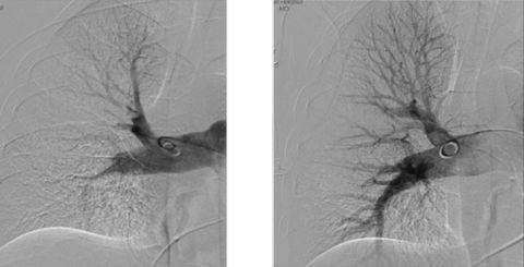
Catheter directed treatment of an acute PE. The first picture shows a clot in the pulmonary artery which does not allow the dye in the blood to pass into the lung. After catheter-directed therapy, dye easily flows through the previously blocked blood vessel.
Pre- and Post-Treatment
IVC Filter Removal
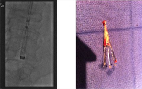
The first picture shows the filter being captured inside the body, and the second picture shows the filter on the surgical table after it has been removed.
Chronic Venous Disease
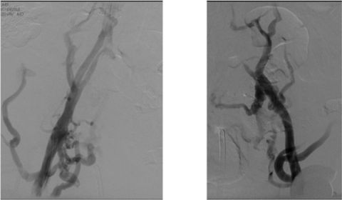
Angioplasty and stenting in a patient with post-thrombotic syndrome. In this patient with very old clots, in the pre-treatment photos, the dye injected through the blood is having a difficult time going through the native vein and has to find alternative pathways to get back to the heart. After angioplasty and stenting (post-treatment photo), the dye easily goes through the newly placed stents. As can be seen from the photographs, the patient’s leg markedly improved.
Pre Treatment
Post Treatment
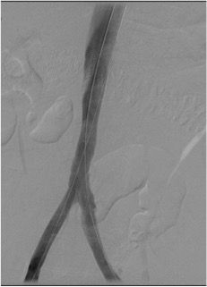
Chronic Venous Disease Treatment pre and post
